
A pregnancy ultrasound is an essential tool for monitoring the health and development of both the mother and the baby during pregnancy. This non-invasive imaging technique uses sound waves to create detailed images of the baby inside the womb, allowing healthcare providers to check on the baby’s growth, position, and overall well-being.
In this article, we’ll walk you through what happens during a pregnancy ultrasound, what to expect, and why this scan is so important for a healthy pregnancy.
What is a Pregnancy Ultrasound?
A pregnancy ultrasound, often referred to as a sonogram, uses high-frequency sound waves to create real-time images of the baby inside the uterus. This non-invasive procedure is commonly performed during various stages of pregnancy to monitor fetal growth and check for any abnormalities. Ultrasound is a crucial tool that helps healthcare providers ensure the health of both the baby and the mother.
At PJ Polyclinic in Petaling Jaya, we use advanced ultrasound services to provide expectant mothers with detailed images and information about their pregnancy. Whether it’s your first trimester or third, an ultrasound provides vital insights into your baby’s development, from confirming the pregnancy to assessing organ growth and movement.
When is a Pregnancy Ultrasound Performed?
Pregnancy ultrasounds are typically performed at key stages during pregnancy to gather important information and ensure that everything is progressing normally. The timing and frequency of ultrasounds depend on various factors, including your health and any risk factors associated with your pregnancy. Here’s when ultrasounds are commonly performed:
- Early Pregnancy Ultrasound (6-8 weeks):
During the early stages of pregnancy, an ultrasound is used to confirm the pregnancy, check the number of embryos, and verify the heartbeats of the baby. This scan can help rule out ectopic pregnancies (where the embryo implants outside the uterus). - Mid-pregnancy Ultrasound (18-22 weeks):
A detailed ultrasound is typically performed between the 18th and 22nd week to assess fetal growth, check for any birth defects, and measure the baby’s organs. It can also determine the position of the placenta and check the amount of amniotic fluid around the baby. - Third Trimester Ultrasound (28-40 weeks):
A third-trimester ultrasound is usually performed if there are concerns about the baby’s growth, position, or fluid levels. It can also be used to assess the baby’s readiness for delivery, such as checking for a breech presentation or assessing the umbilical cord.
What to Expect During Your Pregnancy Ultrasound
During a pregnancy ultrasound, the procedure is generally straightforward and painless, though some variations exist depending on the type of ultrasound being performed. Here’s what you can expect during the scan:
- Preparation
For early ultrasounds, especially in the first trimester, you may be asked to have a full bladder before the exam. This helps provide clearer images, particularly in transabdominal ultrasounds. For later-stage ultrasounds, there’s generally no need for preparation. However, your healthcare provider will give you specific instructions based on your unique situation. - The Procedure
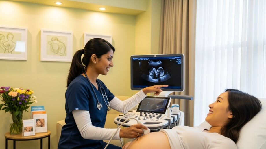
The procedure typically takes around 20-30 minutes. During the ultrasound, you will lie down on an examination table, and a gel will be applied to your abdomen. The gel is used to help transmit the sound waves. The sonographer (the technician performing the scan) will then move a handheld device called a transducer over your abdomen to capture images of the baby and the surrounding structures. In some cases, if the baby’s position makes it hard to see the details through the abdominal scan, a transvaginal ultrasound may be used. This is especially common in early pregnancy ultrasounds when the baby is still small. - Real-time Imaging
Ultrasound provides real-time images, so you’ll be able to see your baby’s movements, heartbeats, and development on the monitor. It’s common to see your baby’s limbs moving, the heart beating, or even the baby sucking their thumb. For many expectant parents, this is an exciting and emotional experience. - After the Scan
Once the ultrasound is complete, the sonographer will clean off the gel, and you can usually return to your normal activities. In some cases, the technician may share a few initial observations, but the final report will typically be sent to your doctor. Your doctor will discuss the findings with you at your next appointment, or if needed, provide further guidance.
Interpreting Ultrasound Results and Next Steps
After your pregnancy ultrasound, the images captured during the scan will be reviewed by a doctor or specialist. Ultrasound results provide valuable information that helps healthcare providers assess both the baby’s development and the mother’s health. Here’s how the results are typically interpreted:
- Fetal Growth and Development
One of the primary goals of a pregnancy ultrasound is to ensure that the baby is growing appropriately. Your doctor will look at various factors, including the size of the baby, the development of organs like the brain and heart, and the baby’s overall movement. Ultrasound measurements can give a clear indication of whether the pregnancy is progressing normally or if further investigation is needed. - Placenta Position and Amniotic Fluid
The ultrasound also provides information about the position of the placenta, which is essential for a safe delivery. Placental issues, such as low-lying or accreta placentas, can pose risks during childbirth. The amount of amniotic fluid around the baby will also be assessed to ensure that the baby has an optimal environment for growth. Any abnormalities in the fluid levels will be monitored and addressed by your healthcare provider. - Detection of Abnormalities
While most pregnancies progress smoothly, ultrasound scans can detect certain abnormalities. These might include congenital conditions, structural problems, or genetic issues. If a potential issue is identified, your doctor may recommend additional testing or follow-up ultrasounds to monitor the situation. Early detection allows for the development of a treatment plan or necessary medical interventions. - Gender Determination
In many cases, ultrasound can determine the sex of the baby, typically around 18-22 weeks of pregnancy. While gender determination is not always 100% accurate, it can provide expectant parents with the option of knowing whether they’re having a boy or a girl, depending on their preferences.
When to Schedule Follow-Up Ultrasounds
In most cases, the routine pregnancy ultrasound between 18-22 weeks is sufficient for monitoring the baby’s health. However, depending on individual circumstances, your healthcare provider may recommend additional ultrasounds throughout the pregnancy, particularly if there are concerns about fetal growth, maternal health, or potential complications.
At PJ Polyclinic in Petaling Jaya, we are here to support you throughout your pregnancy with advanced ultrasound imaging, ensuring that you and your baby receive the best possible care.
FAQ
When is the best time to have a pregnancy ultrasound?
The ideal time for a routine pregnancy ultrasound is between 18 and 22 weeks of gestation. This scan allows for a detailed assessment of the baby’s growth, position, and development. However, ultrasounds can be performed earlier (around 6-8 weeks) to confirm pregnancy and detect the heartbeat, and later in pregnancy if there are concerns or complications.
Is ultrasound safe for both the mother and baby?
Yes, ultrasound is completely safe for both the mother and baby. It uses sound waves instead of radiation, which makes it a non-invasive and harmless method of monitoring pregnancy. Ultrasound is one of the most commonly used imaging tools throughout pregnancy because of its safety.
Can I find out the gender of my baby through an ultrasound?
Yes, most ultrasounds performed during the second trimester (around 18-22 weeks) can reveal the baby’s gender. However, it’s important to note that while ultrasounds are generally accurate, they are not always 100% reliable in determining gender, especially if the baby is positioned in a way that makes it difficult to see.
What happens if something abnormal is found during the ultrasound?
If any abnormalities are detected during the ultrasound, your doctor will discuss the findings with you and recommend further tests or monitoring. Early detection of issues allows for timely interventions and better planning for the pregnancy and delivery.
What should I do to prepare for my pregnancy ultrasound?
For most pregnancy ultrasounds, no special preparation is required. However, for early ultrasounds (especially in the first trimester), you may be asked to have a full bladder, as this helps provide clearer images. Your doctor will provide specific instructions based on the type of ultrasound you need.
The Importance of Pregnancy Ultrasound for a Healthy Pregnancy
Pregnancy ultrasounds are an invaluable tool for monitoring the health and development of both the baby and the mother. From early confirmation of pregnancy to detailed assessments of fetal growth and position, ultrasound provides essential information that helps ensure a healthy pregnancy and a safe delivery.
At PJ Polyclinic in Petaling Jaya, we provide advanced ultrasound services to give expectant mothers peace of mind and comprehensive care. Our experienced team is dedicated to offering you the best possible care throughout your pregnancy journey.
Book Your Ultrasound Today and Ensure Your Baby’s Health
If you’re expecting and would like to schedule your pregnancy ultrasound, book an appointment at PJ Polyclinic in Petaling Jaya today. We are here to support you every step of the way with expert care and the latest ultrasound technology.